| Target lesion | Baseline | Current | Change |
|---|---|---|---|
| Right breast tumour | 33 mm | 20 mm | −39% |
| Liver metastasis (segm. 8) | 45 mm | 29 mm | −36% |
| Right-lung lesion | 13 mm | 8 mm | −38% |
| Mediastinal node (short axis) | 16 mm | 9 mm | −44% |
| Sum of diameters (SLD) | 121 mm | 76 mm | −37.2% |
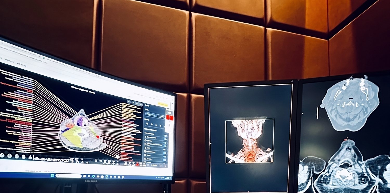
We are about to launch expert CT teleradiology for oncology — drug programmes, monitoring of chemo-, immuno- and radiotherapy response, and clinical trials, in line with RECIST 1.1 and iRECIST criteria. We are already building the team and forming partnerships.
| Target lesion | Baseline | Current |
|---|---|---|
| Right breast tumour | 33 mm | 20 mm |
| Liver metastasis (segm. 8) | 45 mm | 29 mm |
| Sum of diameters (SLD) | 121 mm | 76 mm |
RECIST Oncologic Teleradiology brings together radiologists who have worked in oncologic imaging for over two decades. We are not a general-purpose teleradiology service — we have deliberately narrowed our scope to what we do best.
We report computed tomography only — for oncologic drug programmes and for monitoring the efficacy of chemotherapy, immunotherapy and radiotherapy. Every study is assessed in full clinical context, compared against all of the patient’s available imaging: prior CT, MRI, PET, SPECT and more.
Our radiologists have taken part in international clinical trials and understand the requirements of sponsors and CROs as well as the realities of drug-programme reimbursement. We know that the decision to continue therapy hinges on the precision of a target-lesion measurement. see how we show this in a report →
We report CT — we compare against everything available.
This is not an ordinary study result. It is a structured document with a RECIST 1.1 measurement table, a calculated response category and conclusions ready for drug-programme documentation and tumour boards. See what it looks like in practice — below is an excerpt of a real report structure.
| Target lesion | Baseline | Current | Change |
|---|---|---|---|
| Right breast tumour | 33 mm | 20 mm | −39% |
| Liver metastasis (segm. 8) | 45 mm | 29 mm | −36% |
| Right-lung lesion | 13 mm | 8 mm | −38% |
| Mediastinal node (short axis) | 16 mm | 9 mm | −44% |
| Sum of diameters (SLD) | 121 mm | 76 mm | −37.2% |
Four complete examples — baseline, response assessment, a benign result and a complete response. Each with a RECIST table and conclusions:
5 target lesions, sum of diameters 121 mm as the reference point for later assessments.
Same methodology, same comparison — an unambiguous partial-response category.
When RECIST does not apply — because there is no malignancy. We state that clearly too.
Lymph nodes below the normal threshold — a complete response to treatment per RECIST 1.1.
View all sample reportsFrom drug-programme eligibility to final response assessment — we deliver reads you can base treatment and reimbursement decisions on.
CT reads that meet the requirements of reimbursed drug programmes — complete, on time and prepared around eligibility and monitoring criteria.
Monitoring of chemotherapy, immunotherapy and radiotherapy efficacy against recognised criteria — with an unambiguous response category (CR / PR / SD / PD).
Imaging support for research sites, sponsors and CROs. Our radiologists have worked in international clinical trials and know the rigour of the protocols.
A fresh expert read of CT studies in doubtful, discordant or treatment-critical cases — including a review of prior measurements.
In oncologic response assessment two radiologists can both measure correctly — and reach opposite conclusions. We know these mechanisms from practice, which is why our rule is continuity of one team, one methodology and full multimodal comparison.
The same tumour cut in a different plane yields a different longest diameter. Both radiologists measure correctly, yet the conclusions are opposite: −21% (response) for one, +23% (progression) for the other. Without a continuous measurement baseline the numbers stop being comparable.
A lesion “grows” because immune cells infiltrate it — the treatment is just starting to work. Assessed by RECIST 1.1 alone it would be called progression; iRECIST requires confirmation on the next scan before therapy is stopped.
An osteolytic lesion can be barely visible at first. As the bone heals it sclerotises and “reveals itself” on imaging — looking like a new lesion, though it is proof of treatment response. Without comparison against the full imaging history the error is easy to make.
We expand on each of these mechanisms — with a full explanation, worked numbers and how we prevent them. See also the model report structure that counters these pitfalls.
Read about assessment pitfalls Sample reports“The decision to continue therapy hinges on the precision of a target-lesion measurement. That is why we measure as if a life depended on it — because it does.”
The RECIST Oncologic Teleradiology team
Soon we will work with oncology centres, clinical-trial organisations and other teleradiology companies. Choose your model and let’s agree terms before go-live.
We take over the CT reads generated within drug programmes and oncologic treatment monitoring — relieving your radiology department while guaranteeing a uniform standard of assessment.
We provide radiological assessment of CT studies in clinical trials — from site reads to consistent serial assessments of patients across protocol time points.
We are the expert back-end for broad-profile teleradiology companies. Send us the oncologic CT from drug programmes — we provide the specialist assessment, you keep the client relationship.
From study upload to finished report — with no friction on your side.
DICOM transfer over an encrypted channel together with prior comparison studies (CT, MRI, PET, SPECT) and the necessary clinical context.
The radiologist assesses the study against the patient’s entire imaging history, maintaining continuity of the existing measurement baseline.
A report with a target / non-target lesion table, measurements, sum of diameters and an unambiguous response category per RECIST 1.1 / iRECIST. see the template →
The report within the agreed SLA. In ambiguous cases — direct contact with the reporting radiologist.
We are looking for experienced oncologic radiologists who want to work in a narrow specialty at the highest level. We are building the team and happy to talk already now.
We are looking for physicians for whom oncologic response assessment is a daily craft — not an add-on to on-call shifts.
We go live soon. Write now — we’ll share a sample report structure → and agree terms of collaboration ready for launch, tailored to your organisation. We reply within one business day.